
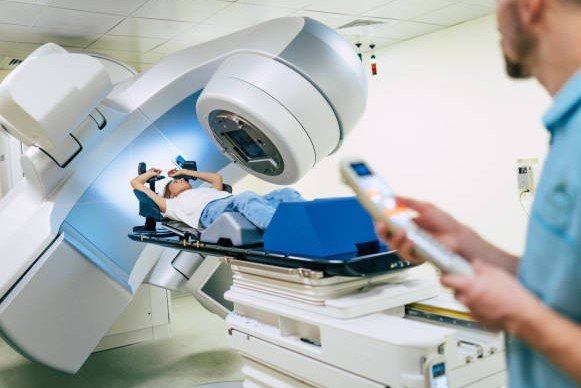
Researchers at the University of Arizona are developing a groundbreaking imaging technology that promises to detect non-melanoma skin cancers more effectively without invasive procedures. Set to begin work in late 2025, this synthetic wavelength imaging approach aims to provide clearer, deeper views into tissues, helping doctors assess tumor invasion and track treatment progress.
What Is Synthetic Wavelength Imaging?
This new method uses two separate light wavelengths to create a virtual, longer wavelength that penetrates deeper into the skin. Unlike traditional tools, it resists light scattering, which often blurs images in deeper tissues.
Experts say this could change how non-melanoma skin cancers like basal cell carcinoma and squamous cell carcinoma are diagnosed. These cancers make up most skin cancer cases, with over five million diagnoses in the United States each year. The technology combines high contrast from shorter wavelengths with better depth from the synthetic one.
The project focuses on challenges unique to these cancers, such as varying lesion sizes and invasion patterns. By tuning the imaging, doctors can get reliable views for both initial checks and ongoing monitoring.
How the Funding Drives Progress
The University of Arizona team secured a 2.7 million dollar grant from the National Institutes of Health to advance this work. Led by optical sciences professor Florian Willomitzer and dermatology expert Dr. Clara Curiel-Lewandrowski, the group plans to build a portable prototype for human testing.
This funding ties into a broader push for non-invasive imaging in biological systems. It addresses gaps in current methods, where tools like confocal microscopy excel at surface details but fail deeper down.
- Grant amount: 2.7 million dollars from NIH.
- Key focus: Non-melanoma skin cancers.
- Timeline: Prototype development starting late 2025.
Researchers aim to make the device versatile, allowing real-time adjustments for different tumor types. This could reduce the need for biopsies in some cases, speeding up care.
The initiative builds on recent trends in medical imaging, where innovations like AI-powered tools have improved early detection rates by up to 20 percent in similar fields.
Challenges in Current Skin Cancer Detection
Traditional imaging faces limits with light scattering in tissues. Visible and near-infrared wavelengths provide good resolution near the surface but lose clarity deeper in.
Longer wavelength options, such as ultrasound, reach further but often lack the detail needed for precise cancer mapping. This gap is critical for non-melanoma cases, which can invade irregularly and require exact margin assessments.
Patients often deal with lesions that vary in depth and spread, making accurate diagnosis tricky. Without better tools, treatments might miss hidden extensions, leading to higher recurrence risks.
In 2025, skin cancer rates continue to rise, driven by factors like increased sun exposure and aging populations. Global data shows non-melanoma skin cancer affects millions, with mortality low but treatment costs high if not caught early.
Potential Impact on Patient Care
This technology could transform how doctors handle skin cancer. By offering tunable depth and resolution, it helps map tumors accurately at diagnosis and during therapy.
For example, monitoring treatment response becomes easier, as the imaging tracks changes without repeated invasive checks. This is vital for non-melanoma types, which account for about 90 percent of all skin cancers.
| Aspect | Current Methods | New SWI Tech |
|---|---|---|
| Depth Penetration | Limited by scattering | Improved with synthetic wavelengths |
| Resolution | High at surface, drops deeper | Balanced high contrast and depth |
| Invasiveness | Often requires biopsies | Fully non-invasive |
| Versatility | Fixed for specific depths | Tunable for various lesions |
Early detection could boost five-year survival rates, already over 95 percent for these cancers when found soon. The portable design means it could reach clinics worldwide, not just major centers.
Integrating this with AI could further enhance accuracy, aligning with 2025’s surge in AI dermatology tools that analyze lesions in real time.
Broader Applications and Future Outlook
Beyond skin cancer, synthetic wavelength imaging might apply to other deep-tissue needs, like monitoring internal organs or vascular issues. Its non-invasive nature opens doors for routine screenings.
Researchers plan initial human studies soon, focusing on safety and effectiveness. If successful, it could roll out in hospitals by the late 2020s.
This fits into global health trends, where non-invasive tech is key to handling rising cancer burdens. Recent events, like the 2025 World Health Organization report on cancer prevention, highlight the need for such innovations to cut diagnosis times.
As work progresses, experts watch closely for how it might reduce healthcare costs, estimated at billions annually for skin cancer treatments.
What do you think about this new imaging tech? Share your thoughts in the comments and spread the word to raise awareness about skin cancer detection.